November 8th is World Radiology Day, a date that invites us to reflect on one of the most important advances in the history of medicine: the discovery of X-rays.
Have you ever wondered how these images that allow us to see inside our body without surgery were discovered? The answer goes back to 1895 and to the German engineer and physicist Wilhelm Conrad Röntgen.
The discovery that changed everything
Röntgen was carrying out experiments with the fluorescence caused by cathode rays that hit certain materials, when almost accidentally, he noticed that an unknown radiation could pass through solid objects such as paper and leave a mark on photographic films. This phenomenon intrigued him so much that he decided to ask his wife, Anna Berta Ludwig, for help.
In this investigation, Röntgen used his partner's hand to capture an image of her bones. That was the first step towards modern X-rays! Röntgen, with great humility, never patented this incredible discovery, allowing the use of X-rays to spread rapidly to hospitals and clinics around the world. In recognition of his contribution to science, he was awarded the Nobel Prize in Physics in 1901.
The most important advances in medical radiology since his discovery allow for faster and more effective treatments:
- Fluoroscopy (1900) Soon after the discovery of X-rays, fluoroscopy allowed real-time observation of internal body movements. This technique is still useful in guided procedures such as cardiac catheterizations and stent placement.
- Computed Tomography (CT) (1970) The invention of computed tomography (CT) by Godfrey Hounsfield and Allan Cormack in the 1970s was a fundamental advance. It uses X-rays to create detailed cross-sectional images of the body, providing a much clearer view of organs and soft tissues compared to traditional X-rays.
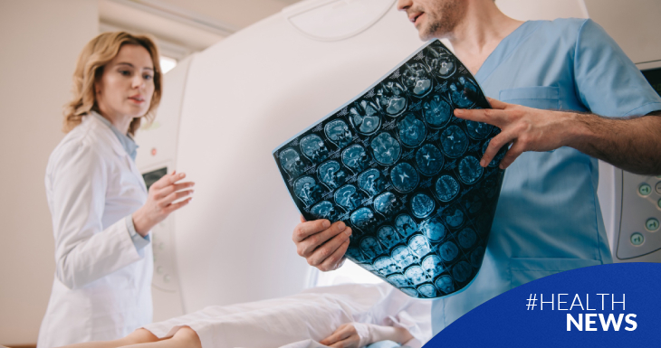
- Magnetic Resonance Imaging (MRI) (1980) In the early 1980s, magnetic resonance imaging (MRI) was introduced as a noninvasive technique that uses magnetic fields and radio waves to obtain detailed images of the body's organs and soft tissues. This technique has been vital in the diagnosis of brain and cardiovascular diseases, and in musculoskeletal studies.
- Ultrasound Although it began in the 1950s, the medical use of ultrasound has advanced considerably in recent decades. This noninvasive technique, which uses sound waves to generate images of the inside of the body, is essential in monitoring pregnancy, diagnosing abdominal conditions, and evaluating the heart.
- Positron Emission Tomography (PET) (1990) PET is an advanced nuclear imaging technique that provides information about cellular metabolism. It is especially useful for detecting cancer and studying the brain, as it can show how tissues are functioning at the cellular level.
- Interventional Radiology This field has evolved to allow radiologists to perform minimally invasive image-guided procedures, such as catheter insertion, biopsies, and tumor treatments using embolization, reducing the need for more invasive surgeries.
- Artificial Intelligence in Radiology In recent years, artificial intelligence (AI) has begun to play a key role in the interpretation of radiological images. Algorithms can detect patterns that humans might miss, which improves diagnostic accuracy and helps radiologists process large volumes of images more quickly.
The importance of radiology in health
By using imaging techniques, detailed and precise information is obtained about the state of the organs and tissues of the human body. One of the main advantages is its ability to detect diseases in early stages, which allows timely treatment to be started and significantly improves the chances of successful patients recovery.
Among the most well-known specific applications we can mention:
- Identifying fractures
- Observing the development of fetuses
- Neurological diagnoses
- Knowing the dental state of the patient
- Locating anomalies
- Detecting signs of arthritis
- Detecting possible types of cancer, especially bone cancer
The risks to which radiologists are subjected:
When we undergo a radiological examination, we are always given instructions to avoid exposure to X-rays as much as possible. But what about the radiological risks to which radiologists are subjected in the imaging room? The main problems that can be faced are: developing some type of cancer, infertility, blindness, cardiovascular diseases, kidney problems, and hair loss. To avoid these risks, the professional must wear a special suit or work in a shielded booth, and their work schedule must provide flexible hours in which they can take a prudent amount of time off, so as not to be overexposed to radiation.
What happens to patients who undergo this type of study?
Different diagnostic tests require different amounts of radiation, but most use low doses that are generally considered safe. However, radiation exposure is cumulative, regardless of the interval between tests. This means that if the person undergoes many diagnostic tests that use low doses or several tests that use high doses, they may be exposed to relatively significant amounts of radiation. The higher the cumulative dose, the greater the risk of cancer and, in some cases, tissue damage.
When ordering diagnostic tests, doctors should consider a person's total exposure to radiation based on their medical history and follow these guidelines based on this:
- Use tests that do not require radiation, such as ultrasound or magnetic resonance imaging (MRI), whenever possible
- Recommend diagnostic tests that use particularly high doses of radiation (such as computed tomography [CT] scans) only when these tests are strictly necessary
- Take precautions to limit radiation exposure during tests (for example, by shielding vulnerable parts of the body, such as the thyroid gland or the abdomen of a pregnant woman), whenever possible
The following table shows the different doses of radiation received in different tests:
| Test Type | Approximate number of chest X-rays needed to receive the same dose | Approximate environmental exposure equivalent to dose † |
|---|---|---|
| Single view of a chest X-ray (from the back to the front) | 1 | 10 days |
| Mammography | 10 | 25 days |
| Lumbar spine X-ray series | 75 | 180 days |
| Cranial CT scan | 100 | 243 days |
| CT scan of the abdomen/pelvis | 300–400 | 2–2,7 years |
| Angiography of the coronary arteries during cardiac catheterization | 350–750 | 2,3-4,9 years |
* These doses represent how much radiation is received and the degree of susceptibility to radiation damage of the exposed body part.
† People are constantly exposed to low levels of naturally occurring radiation, but the amount of radiation varies depending on different geographic locations
CT = computed tomography
Reference: American College of Radiology(ACR): Radiation Dose to Adults From Common Imaging Examinations
SOURCE: www.merckmanuals.com
Radiology Day highlights the crucial role of medical imaging in modern medicine: from simple bone X-rays to advanced MRIs, radiology makes it possible to diagnose diseases, monitor the progress of treatments and make more informed medical decisions.
